BIOSCIENCE CORES
  • Home
  • Proteomics
    • Rates
    • Resources
    • Protocols
  • Imaging
    • Imaging Core >
      • Instruments >
        • Zeiss LSM 980
        • Leica SP5X
        • Zeiss LSM710
        • DeltaVision
        • Nikon Eclipse
        • Zeiss AxioObserver
      • NEW USER GUIDE
      • RATES
      • FAQ
      • IMAGE GALLERY
    • Imaging Incubator >
      • INSTRUMENTS >
        • PerkinElmer
        • ASI diSPIM
        • JPK AFM
        • 3P
      • Rates
      • NEW USER GUIDE
  • Flow Cytometry
  • Genomics
    • Covaris S220
    • Agilent TapeStation

Imaging Incubator

INCUBATOR HOME
INSTRUMENTS
RATES
NEW USER GUIDE

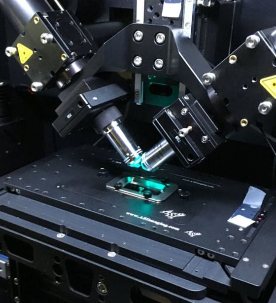
ASI diSPIM Lightsheet

Picture
  • The ASI di-SPIM (dual-view Selective Plane Illumination Microscope) allows for rapid and gentle imaging of fluorescently labeled samples mounted on a chambered coverglass (in liquid) and placed on an inverted microscope XY stage.
  • The method  uses two objective lenses placed at right angle in order to illuminate a thin slice of the sample perpendicular to the direction of the observation. Because only the focus plane of the sample is illuminated,  photobleaching and photoxicity are minimized, making it an ideal method for imaging living cells and small groups of cells/embryos (up to 300 um sample depth) such as C. elegans
  • The di-SPIM is capable of rapid volumetric imaging: up to 10 volumes per second, and (computationally-generated) isotropic resolution (<400nm with 40x 0.8NA objective, others available).
  • A chamber with temperature and humidity control is available for live mammalian samples.
  • Excitation wavelengths include: 405, 488, 561, 640nm

New Users

Faculty and students who wish to use the microscope should follow the instructions in the New Users Guide.

Policy

The microscope is available to trained users on an equal basis. For a comprehensive list of policies (including BSL-2 protocols), please see the Facility SOP.

System Specifications

Microscope Stand
Inverted MIM (modular infinity microscope)
Illumination
405, 488, 561, 640nm lasers
Emission
Long-pass emission filters available for each laser
Light detectors
Two ZYLA 4.2 sCMOS dual-Camera Link cameras,
bottom USB2 CCD camera for epi-fluorescence and TL observation
Objective Lenses
10x, 20x, 40x (all water dipping lenses)
Incubation
Chamber available for temperature and CO2 control
X-Y Stage
Motorized
Sample chamber
Fits only the following size cover glass: 24 x 50mm, #1.5
Software
Micromanager 1.4

References

  • Imaging Incubator Policy
  • ASI website

Contact Us

Dr. Aleksandr Smirnov
Associate Research Scientist
Institute for Physical Sciences  & Technology (IPST)
University of Maryland, College Park
asmirnov@umd.edu

Picture

HOMe
biological sciences home
cmns home
cbmg home
web accessibility
  • Home
  • Proteomics
    • Rates
    • Resources
    • Protocols
  • Imaging
    • Imaging Core >
      • Instruments >
        • Zeiss LSM 980
        • Leica SP5X
        • Zeiss LSM710
        • DeltaVision
        • Nikon Eclipse
        • Zeiss AxioObserver
      • NEW USER GUIDE
      • RATES
      • FAQ
      • IMAGE GALLERY
    • Imaging Incubator >
      • INSTRUMENTS >
        • PerkinElmer
        • ASI diSPIM
        • JPK AFM
        • 3P
      • Rates
      • NEW USER GUIDE
  • Flow Cytometry
  • Genomics
    • Covaris S220
    • Agilent TapeStation