Frequently Asked Questions
1. What thickness coverslip should I use?
2. But what if I need to image cells in a 6-, 12-, 96-well plate with a plastic bottom?
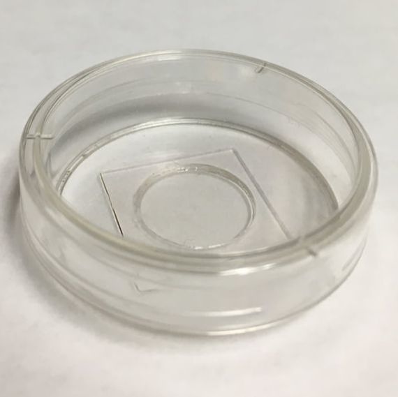
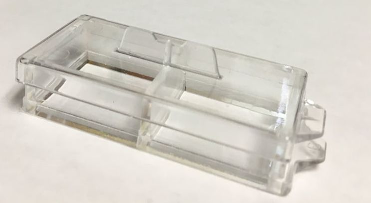
3. What if I want image live samples on an inverted scope and/or I don’t want to mount my sample on a slide?
- 0.17mm (#1.5) coverslip/dish/chamber slide unless you have a good reason not to, and can explain that reason.
2. But what if I need to image cells in a 6-, 12-, 96-well plate with a plastic bottom?
- Use the Zeiss AxioObserver inverted fluorescence microscope. It has a plate adapter and long working distance lenses (20x, 40x) that are capable of imaging through plastic. You will need to adjust the microscope objective correction collar (please see the director for help with this). Don’t expect high resolution images. If you need higher quality images, use a 35mm dish or chambered coverglass with a #1.5 coverslip as described below.
3. What if I want image live samples on an inverted scope and/or I don’t want to mount my sample on a slide?
- Use a 35mm dish or chambered coverglass with a #1.5 coverslip. A number of companies sell these. Examples include: 1) MatTek 35mm Glass Bottom microwell dish, part number, P35G-1.5-10-C, 2) Lab-Tek II Chambered #1.5 Coverglass System (single chamber #155360; 2 chamber #155379; 4 chamber #155382, 8 chamber #155409.
4. Why do I want to use a confocal (other than the fact that my PI told me to?)
5. What dye combinations should I use?
6. What if I want to take a color image of a non-fluorescently labeled sample, like an immunohistochemical stain?
7. What are the main differences between the two confocals?
8. Why would I ever want to use the Zeiss if the Leica has so many more advanced features?
9. Can I image live BSL-2 samples?
4. Why do I want to use a confocal (other than the fact that my PI told me to?)
- Because the confocal pinhole will block light emitted from above and below the focal plane of your sample, resulting in higher quality images, and the ability to image thin optical sections in multiple planes (z-stack). The thicker your sample (up to 100 µm), the more you will benefit from confocal imaging.
5. What dye combinations should I use?
- If you have a choice, make sure your dyes/lesson proteins spectrally separated from each other so that you don’t get cross talk/bleedthrough.
- For example, GFP and YFP together is a terrible combination, because both proteins are excited by the same wavelength of light, and their emission spectra directly overlap. To image these two fluorescent proteins together, you’d need to do spectral imaging on the Leica or Zeiss confocal, and will need to prepare appropriate controls (single labeled samples).
- For multi-labeled samples, a typical combination might look like this: DAPI/Hoescht (blue), GFP/FITC (green), DsRed/rhodamine/Alexa568 (red) and Cy5/Alexa 633 (far red).
6. What if I want to take a color image of a non-fluorescently labeled sample, like an immunohistochemical stain?
- Use the Nikon Ti upright microscope with color camera.
7. What are the main differences between the two confocals?
- The Leia SP5x has a number of advanced features that are lacking on the Zeiss LSM710: a resonance scanner for high-speed imaging, a z-galvo stage for fast z-stacks, a stage-top environmental chamber with heat, CO2, and humidity, a White Light Laser capable of illuminating your sample anywhere between 470-670nm in 1nm increments, a more sensitive HyD detector, and a motorized stage for tile scanning and point visiting.
8. Why would I ever want to use the Zeiss if the Leica has so many more advanced features?
- Because the Zeiss works just fine if you need to take z-stacks of fixed cells and/or tissue (with up to 4 labels), and don't need to tile-scan. Plus, the Zeiss is used a bit less than the Leica, so it is easier to schedule time on the instrument. That said, if your sample is particularly dim, you will benefit from the HyD detector on the Leica.
9. Can I image live BSL-2 samples?
- Yes, under the conditions outlined in the Facility SOP (page 2).
Contact Us
|
Dr. Charles Delwiche
Faculty Supervisor [email protected] 301.405.8286 2108 Bioscience Research Building |


