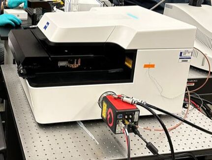
Zeiss Lattice Lightsheet 7
|
|
New Users
Faculty and students who wish to use the microscope should follow the instructions in the New Users Guide. Training takes ~2 to 3 hrs, with additional mandatory follow-up sessions. Users are encouraged to bring their own samples to the training session. If none are available, prepared slides will be used.
Policy
The microscope is available to trained users on an equal basis. An individual user may reserve one four hour block of time during "peak" daytime hours, and an additional 6 "off-peak" hours (all other times). Investigators performing live cell imaging experiments may signup for overnight blocks of time after 6pm.
For a comprehensive list of policies (including BSL-2 protocols), please see the Facility SOP.
For a comprehensive list of policies (including BSL-2 protocols), please see the Facility SOP.
Rates
The rate schedule applies to different users and is current for the 2022/2023 academic year. Fees are used to help cover the cost of the service contract on the microscope, and to help pay for materials such as lens paper, lens cleaning solution and mercury bulbs.
System Specifications
|
References
Imaging Incubator Protocols:Useful Links:
Contact Us
Amy Beaven
CBMG Imaging Core Director
[email protected]
301.405.7238
0107 Microbiology Building
CBMG Imaging Core Director
[email protected]
301.405.7238
0107 Microbiology Building