BIOSCIENCE CORES
  • Home
  • Imaging
    • Imaging Core >
      • Instruments >
        • Zeiss LSM 980
        • Leica Stellaris 8
        • Zeiss LSM710
        • DeltaVision
        • Nikon Eclipse
        • Zeiss AxioObserver
      • NEW USER GUIDE
      • RATES
      • FAQ
      • IMAGE GALLERY
    • Imaging Incubator >
      • INSTRUMENTS >
        • Zeiss LLS7
        • PerkinElmer
        • ASI diSPIM
        • JPK AFM
        • 3P
      • Rates
      • NEW USER GUIDE
  • Brain & Behavior Institute
  • Genomics

Imaging Core

IC HOME
INSTRUMENTS
RATES
NEW USER GUIDE
FAQ
IMAGE GALLERY

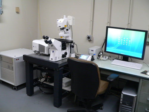
Zeiss LSM710 Laser Scanning Confocal

Picture
  • Ideal for imaging diverse fluorophores in fixed and live samples.
  • Suitable applications include colocalization studies, FRAP, FRET, spectral imaging, and time courses.
  • Lasers: 405nm, argon (458, 488, 514nm), 561nm, 633nm.
  • Objectives:  10x, 20x, 40x oil, 63x oil, 63x water, 100x oil.
  • Features: 3 photomultiplier tubes (PMTs), one transmitted light PMT, manual x-y stage, Zen software.
  • Analysis: An off-line workstation is available for imaging analysis.

New Users

Faculty and students who wish to use the microscope should follow the instructions in the New Users Guide. Training takes ~2 to 3 hrs, with additional mandatory follow-up sessions required until the director determines the user is capable of operating the microscope independently. Users are encouraged to bring their own samples to the training session. If none are available, prepared slides will be used. It is also highly recommended to review the UMD Zeiss LSM710 Quick-Start Guide before training begins.

Policy

The microscope is available to trained users on an equal basis. Individual users may reserve one four hour block of time during "peak" daytime hours, and an additional 6 "off-peak" hours (all other times).

For a comprehensive list of policies (including BSL-2 protocols), please see the Facility SOP.

Rates

The rate schedule applies to all users and is current for the 2022/2023 academic year. Fees are used to help cover the cost of the service contract on the microscope, which is partially subsidized by the department, and to help pay for materials such as lens paper, lens cleaning solution and mercury bulbs.

System Specifications

Microscope Stand
Zeiss AxioObserver inverted stand
Illumination
405 diode, Argon (458, 488, 514nm), 561nm, 633nm
Emission
Fully spectral
Light detector
3 PMTs, 1 transmitted light PMT
Objective Lenses
10x/0.3NA/5.5mm WD
40x/1.3 NA oil/0.2mm WD
63x/1.4NA oil/0.190mm WD
63x/1.2NA water/0.280mm WD
100x/1.4NA oil/0.170mm WD
Incubation
None
X-Y Stage
Manual
Software
Zen

References

Imaging Core protocols:
  • Zeiss LSM710 Startup and Shutdown
  • Zeiss LSM710 Quickstart Guide
  • Zeiss LSM710 Policy

Useful links:
  • Zeiss Homepage
  • Zeiss Zen 2012 Lite software download request page

Contact Us

Amy Beaven
Facility Director
[email protected]
301.405.7238
0107 Microbiology Building

Dr. Charles Delwiche
Faculty Supervisor
[email protected]
301.405.8286
2108 Bioscience Research Building

Picture

HOMe
biological sciences home
cmns home
cbmg home
web accessibility
  • Home
  • Imaging
    • Imaging Core >
      • Instruments >
        • Zeiss LSM 980
        • Leica Stellaris 8
        • Zeiss LSM710
        • DeltaVision
        • Nikon Eclipse
        • Zeiss AxioObserver
      • NEW USER GUIDE
      • RATES
      • FAQ
      • IMAGE GALLERY
    • Imaging Incubator >
      • INSTRUMENTS >
        • Zeiss LLS7
        • PerkinElmer
        • ASI diSPIM
        • JPK AFM
        • 3P
      • Rates
      • NEW USER GUIDE
  • Brain & Behavior Institute
  • Genomics