About
|
The Imaging Core (IC) provides researchers in the Life Sciences with the opportunity to image live, fixed, unlabeled or fluorescently labeled samples using one of several conventional and advanced light microscope systems, including upright and inverted fluorescence microscopes, laser scanning confocal and total internal reflection.
Faculty, post-docs, graduate and undergraduate students with any level of experience, from any department, are invited to use the facility's microscopes. Outside users are also welcome, though priority goes to UMD researchers.
ServicesThe director provides training on all equipment, guidance on experimental design, and assistance with microscope operation.
|
Download a copy of the Imaging Core Brochure.
|
Instruments*
*See the Instruments page for an overview of all scopes, or select an instrument below for detailed information on that scope.
Zeiss LSM 980 Airyscan 2
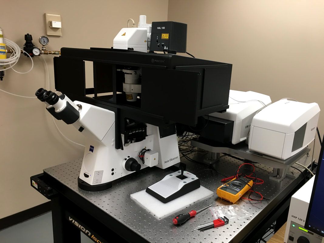
DeltaVision Elite w/TIRF
|
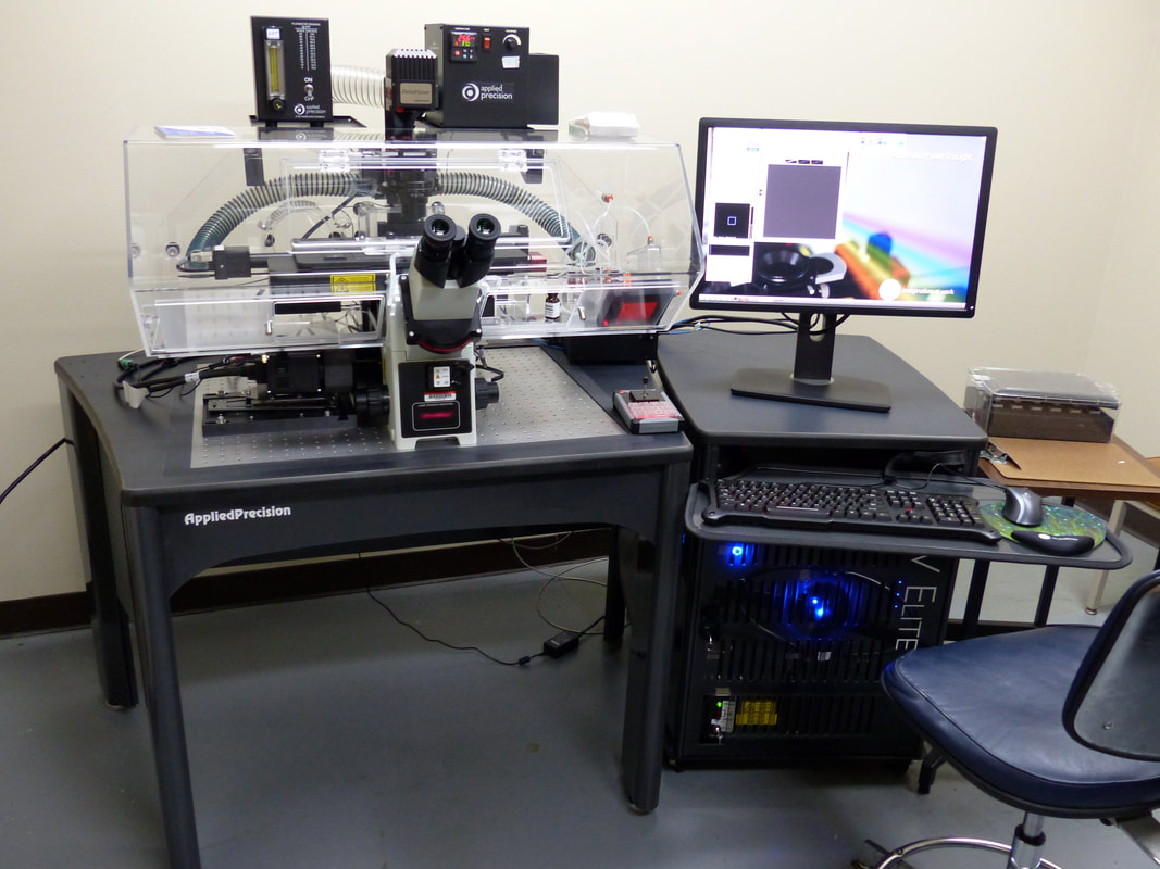
Leica Stellaris 8 FALCON
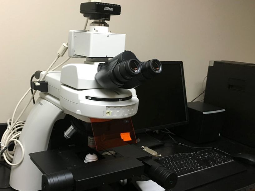
Nikon Eclipse
|
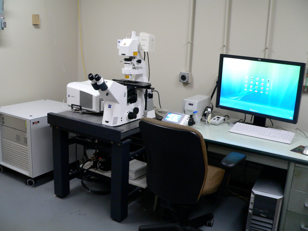
Zeiss LSM710 confocal
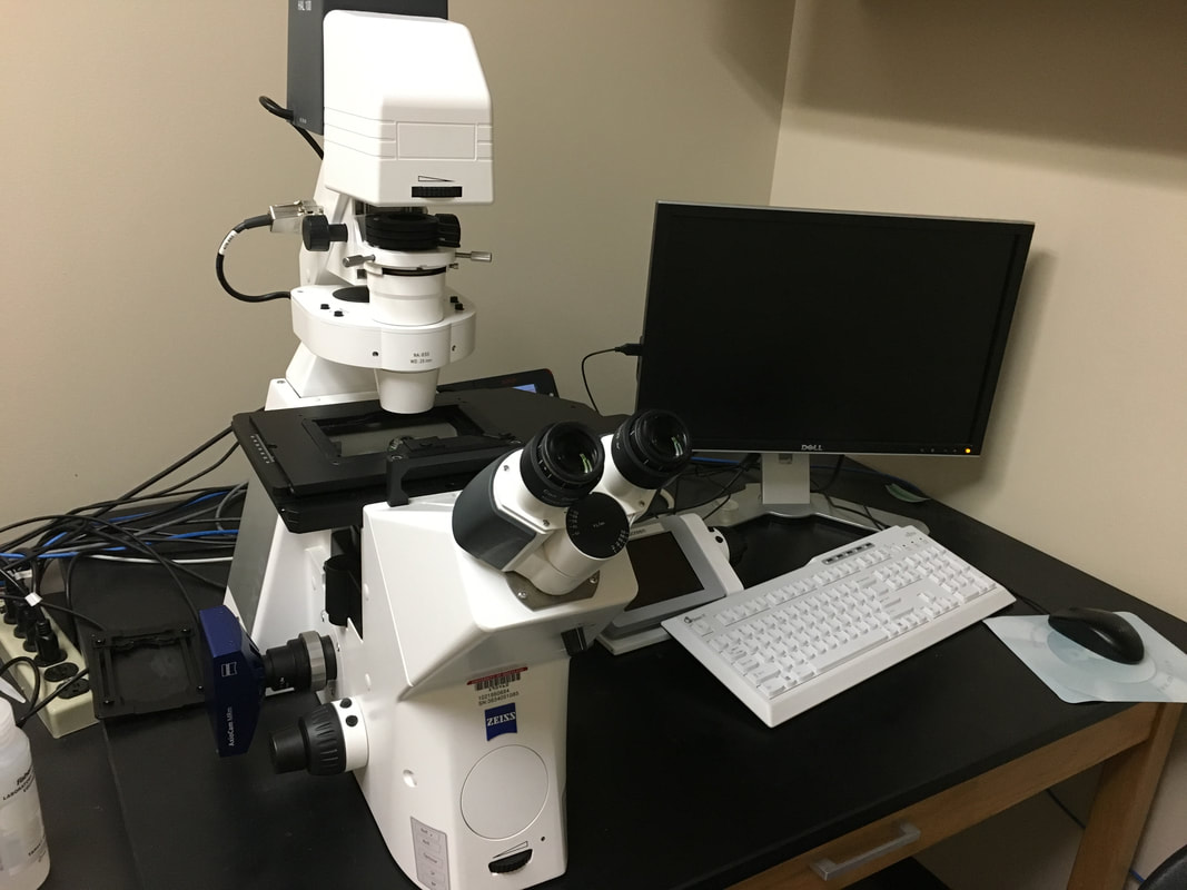
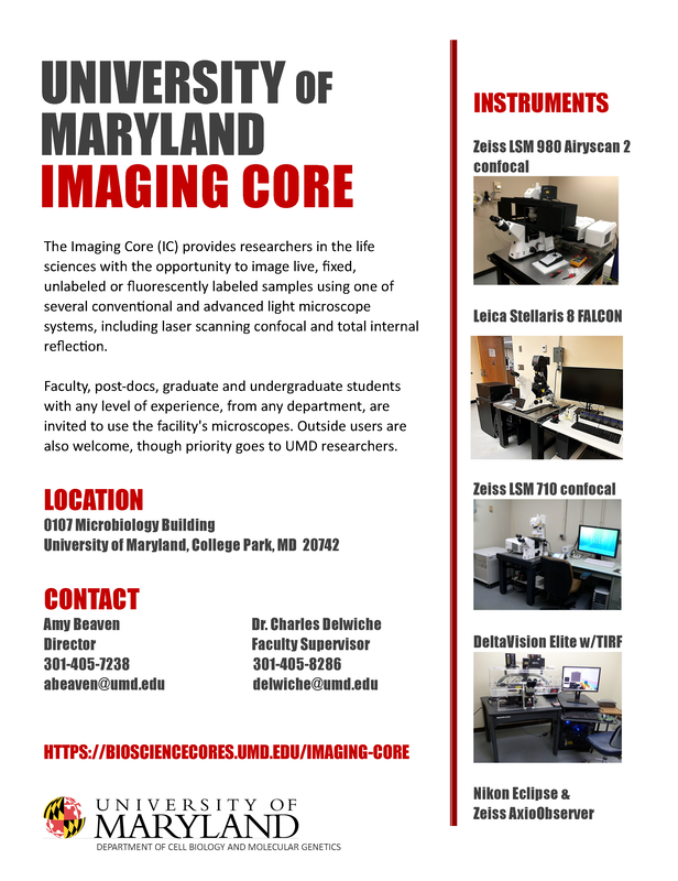
Zeiss AxioObserver
|
Additional instruments (PerkinElmer spinning disk, ASI diSPIM, JPK NanoWizard 4a AFM) are located in the CMNS Imaging Incubator (Physical Sciences Complex building).
Acknowledging the Imaging Core
- It is important to properly acknowledge the use of the Imaging Core in your publications. Acknowledgement helps us demonstrate and document how the Imaging Core contributes to the research community. It also aids us in our efforts to secure more funding to purchase additional instruments and offer new services.
- Suggested wording: "We acknowledge the Imaging Core Facility in the department of Cell Biology and Molecular Genetics at the University of Maryland, College Park for [name of instrument; brief description of assistance].
- Zeiss LSM 980 users: the NIH requires that all publications containing data collected on the Zeiss LSM 980 Airyscan, including press releases, acknowledge NIH grant support with a disclaimer such as the following: Purchase of the Zeiss LSM 980 Airyscan 2 was supported by Award Number 1S10OD025223-01A1 from the National Institute of Health.
- Leica Stellaris 8 users: the NIH requires that all publications containing data collected on th eLeica Stellaris 8, including press releases, acknowledge NIH grant support with a disclaimer such as the following: Purchase of the Leica Stellaris 8 was supported by Award Number 1S10ODO34260 from the National Institute of Health.
- Please send a PDF copy of all publications to the director, Amy Beaven.
Contact Us

|
Amy Beaven
CBMG Imaging Core Director [email protected] 301.405.7238 0107 Microbiology Building |
Dr. Charles Delwiche
Imaging Facilities Supervisor [email protected] 301.405.8286 2108 Bioscience Research Building |