DeltaVision Elite Deconvolution/TIRF
|
|
New Users
Faculty and students who wish to use the microscope should follow the instructions in the New Users Guide. Training takes ~2 to 3 hrs, with additional mandatory follow-up sessions required until the director determines the user is capable of operating the microscope independently. Users are encouraged to bring their own samples to the training session. If none are available, prepared slides will be used. It is also highly recommended to review the DeltaVision Quickstart Guide before training begins.
Policy
The microscope is available to trained users on an equal basis. Individual users may reserve one four hour block of time during "peak" daytime hours, and an additional 6 "off-peak" hours (all other times). Investigators performing live cell imaging experiments may signup for 12 hours blocks of time after 6pm.
For a comprehensive list of policies (including BSL-2 protocols), please see the Facility SOP.
For a comprehensive list of policies (including BSL-2 protocols), please see the Facility SOP.
Rates
The rate schedule applies to all users and is current for the 2022/2023 academic year. Fees are used to help cover the cost of the service contract on the microscope, which is partially subsidized by the department, and to help pay for materials such as lens paper, lens cleaning solution and mercury bulbs.
System Details
|
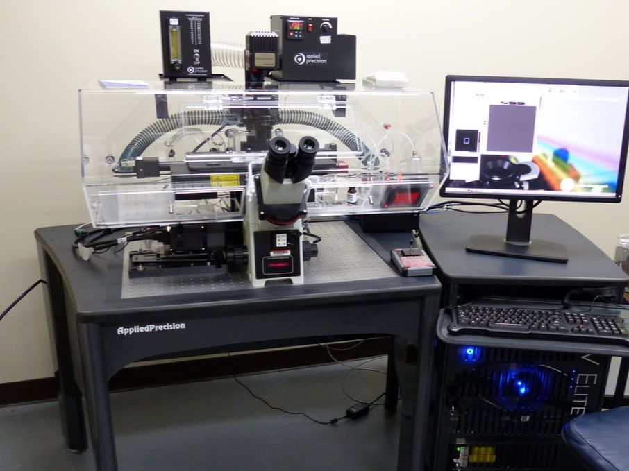
The DeltaVision Elite Deconvolution/TIRF microscope system is a 3-D deconvolution (image restoration) system with 488nm and 561nm laser lines for TIRF (total internal reflection microscopy).
Deconvolution uses the properties of the microscope system (point spread function) to deblur and remap out-of-focus fluorescent signal in order to enhance image resolution and contrast. TIRF is a technique used to image samples within ~100-200nm of the coverslip surface. The system is equipped a number of objective lenses (including 60x oil, 60x water and 60x TIRF), a CMOS camera, and DAPI, CFP, GFP, YFP, TRITC, mCherry, and CY5 fluorescence filters. It is recommended for fluorescence imaging of thin fixed specimens and time-lapse imaging of live cells. The proprietary solid state illumination system provides uniform, high intensity light to the sample, enabling detection of small, dim objects, and allowing for shorter exposure times and fast image acquisition. An incubation system with temperature control and 5% CO2 is available for long-term live cell imaging experiments. The system is equipped with an Ultimate Focus laser that can help prevent z-drift during time-lapse studies. When in TIRF mode (excitation 488, 561nm), the system images samples within 100-200nm of the coverslip surface. The TIRF angle is variable and each wavelength is chromatically corrected to ensure both laser lines penetrate the sample to the same depth.
|
References
Imaging Core protocols:
Contact Us
|
Dr. Charles Delwiche
Faculty Supervisor [email protected] 301.405.8286 2108 Bioscience Research Building |