BIOSCIENCE CORES
  • Home
  • Imaging
    • Imaging Core >
      • Instruments >
        • Zeiss LSM 980
        • Leica Stellaris 8
        • Zeiss LSM710
        • DeltaVision
        • Nikon Eclipse
        • Zeiss AxioObserver
      • NEW USER GUIDE
      • RATES
      • FAQ
      • IMAGE GALLERY
    • Imaging Incubator >
      • INSTRUMENTS >
        • Zeiss LLS7
        • PerkinElmer
        • ASI diSPIM
        • JPK AFM
        • 3P
      • Rates
      • NEW USER GUIDE
  • Flow Cytometry
  • Genomics

Imaging Incubator

INCUBATOR HOME
INSTRUMENTS
RATES
NEW USER GUIDE

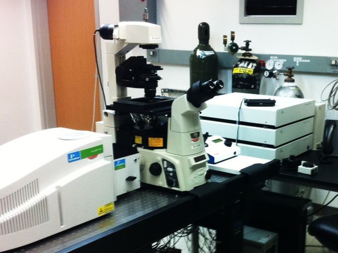
PerkinElmer Spinning Disk Confocal

Picture
  • The PerkinElmer UltraView VoX confocal spinning disk system is optimized for rapid live-cell imaging in four dimensions (x, y, z and time).
  • The system produces high signal-to-noise ratio images with minimal photobleaching.
  • It is equipped with a Yokogawa CSU-X1 spinning disk scanhead (5000 rpm), six solid-state lasers ( 405, 440, 488, 515, 561, 640nm), a 512x512 Hamamatsu EMCCD camera, a Tokai Hit environmental chamber with temperature, humidity, and C02 control, a motorized x-y stage, piezo-z, and a PhotoKinesis unit for FRAP and photo-activation/conversion. 

New Users

Faculty and students who wish to use the microscope should follow the instructions in the New Users Guide. Training takes ~2 to 3 hrs, with additional mandatory follow-up sessions. Users are encouraged to bring their own samples to the training session. If none are available, prepared slides will be used.

Policy

The microscope is available to trained users on an equal basis. An individual user may reserve one four hour block of time during "peak" daytime hours, and an additional 6 "off-peak" hours (all other times). Investigators performing live cell imaging experiments may signup for 12-14 hours blocks of time after 6pm.

For a comprehensive list of policies (including BSL-2 protocols), please see the Facility SOP.

Rates

The rate schedule applies to different users and is current for the 2022/2023 academic year. Fees are used to help cover the cost of the service contract on the microscope, and to help pay for materials such as lens paper, lens cleaning solution and mercury bulbs.

System Specifications

Microscope Stand
Nikon TI inverted
Illumination
Lasers: 405, 440, 488, 514, 561, 640nm
Emission
445w60, 485w60, 525w50, 550w49, 615w70, 705w90
Light detector
512x512 Hamamatsu EMCCD. Up to ~57 frames per second with 1ms exposure. Resolution (microns per pixel): 20x: 0.717, 40x: 0.36, 60x oil: 0.234, 100x: 0.14.
Objective Lenses
10x phase, 20x 0.75NA, 40x 1.3NA oil, 60x 1.4NA oil, 100x 1.49NA oil, 60x 1.2NA water
Incubation
Tokai Hit stage-top incubator with temperature, humidity, CO2 control. Can accommodate 35mm dishes, chambered coverglass, and 3, 6, 12-well plates.
X-Y Stage
Motorized
Software
Volocity Acquisition, Visualization, Quantitation software modules

References

Imaging Incubator Protocols:
  • PerkinElmer Start-Up and Shut-down Procedure
  • PerkinElmer Policy
  • Nikon PFS with Volocity

Useful Links:
  • PerkinElmer

Contact Us

Amy Beaven
CBMG Imaging Core Director
abeaven@umd.edu
301.405.7238
0107 Microbiology Building
Picture

HOMe
biological sciences home
cmns home
cbmg home
web accessibility
  • Home
  • Imaging
    • Imaging Core >
      • Instruments >
        • Zeiss LSM 980
        • Leica Stellaris 8
        • Zeiss LSM710
        • DeltaVision
        • Nikon Eclipse
        • Zeiss AxioObserver
      • NEW USER GUIDE
      • RATES
      • FAQ
      • IMAGE GALLERY
    • Imaging Incubator >
      • INSTRUMENTS >
        • Zeiss LLS7
        • PerkinElmer
        • ASI diSPIM
        • JPK AFM
        • 3P
      • Rates
      • NEW USER GUIDE
  • Flow Cytometry
  • Genomics