Instrument |
Style |
Type |
Objectives |
Sample Type |
Stage |
Incubation |
Relative Speed |
Lasers/ Filters |
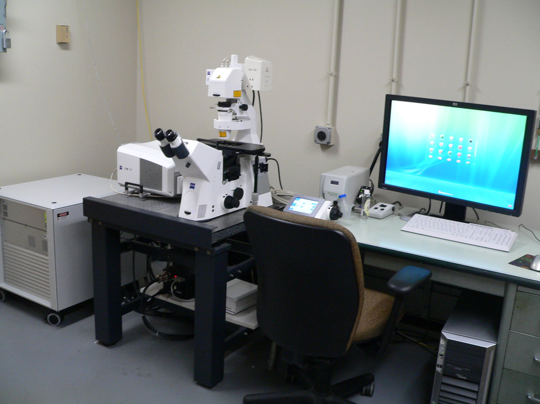
Zeiss LSM710 |
Inverted |
Laser scanning confocal fluorescence |
5, 10, 40x oil, 63x oil, 100x oil |
Live or fixed cells or tissues |
Manual |
None |
Slower |
405, 458, 488, 514, 561, 633nm |
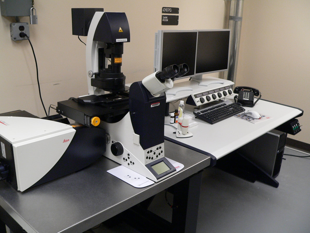
Leica SP5X |
Inverted |
Laser scanning confocal fluorescence |
10, 20, 40x oil, 63x oil, 63x water, 63x glyc |
Live or fixed cells or tissues |
Motorized in x,y,z |
Tokai Hit Stagetop with CO2 |
Slower |
405, argon (458, 488, 514), white light laser (470-670nm) |
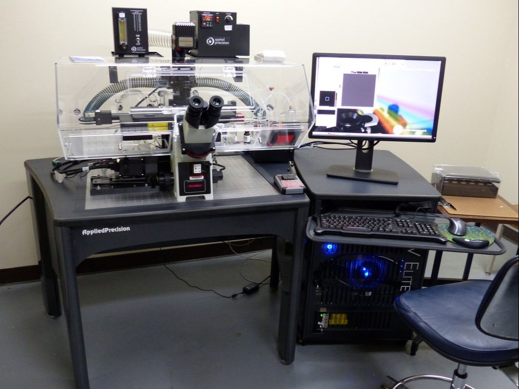
DeltaVision Deconvolution/TIRF |
Inverted |
Deconvolution/ Total Internal Reflection fluorescence |
10x, 20x LWD, 40x LWD, 60x oil, 60x water, 60x TIRF |
Live or fixed cells |
Motorized in x,y,z |
"Big box" Incubator with CO2 |
Faster |
DAPI/GFP/DsRed/A633 or CFP/YFP/mCherry TIRF: 488, 561nm lasers |
Zeiss AxioObserver |
Inverted |
Widefield fluorescence |
10x, 20x LWD, 40x LWD, 100x oil |
Live or fixed cells, suitable for cells in plastic plates |
Manual |
Stagetop Incubator, no CO2 |
Faster |
Standard DAPI, GFP, dsRed, CFP, YFP filters |
Nikon Eclipse |
Upright |
Brightfield with color camera, fluorescence with monochrome camera |
4x, 10x, 20x, 40x oil |
Live or fixed cells or tissue |
Motorized x-y |
None |
Faster |
DAPI, GFP, dsRed (manual switching) |
|
FAQ
|
IMAGE GALLERY
|
For all your light microscopy needs....The Imaging Core (IC) provides researchers with the opportunity to image live, fixed, unlabeled or fluorescently labeled samples using one of several available microscopes. Services include training on all equipment, guidance on experimental design, and technician-assisted microscope operation.
|
|
|
Leica SP5X Laser Scanning Confocal
Ideal for imaging diverse fluorophores in fixed and live samples. Lasers include: 405nm, argon (458, 488, 514nm), and White Light (470-670nm, tunable in 1nm increments). System is equipped with a low-noise hybrid detector, motorized x-y stage, environmental chamber, and resonance scanner for high-speed imaging.
|
Zeiss LSM710 Laser Scanning Confocal
Ideal for imaging fixed, live (room temperature), single or multi-labeled samples. System is equipped with a manual stage, 3 light detectors, and 6 major laser lines (405, 458, 488, 514, 561, 633nm) Suitable applications include colocalization studies, immunofluorescence, FRAP and time courses.
|
|
|
DeltaVision Elite Deconvolution/TIRF
The DeltaVision is a 3-D deconvolution system with 488 and 561nm lasers for TIRF (total internal reflection microscopy). Features include motorized x-y stage, environmental chamber, and epi filters for DAPI, CFP, GFP, YFP, dsRed, mCherry, Cy5. The system excels at imaging "small, dim, live" samples.
|
Zeiss AxioObserver FluorescenceInverted microscope equipped phase contrast, standard fluorescence filters (DAPI, GFP, DsRed, CFP, YFP), and monochrome camera. Suitable for imaging of bacteria and thin fluorescently labeled samples. Two long-working distances lenses (20, 40x) and 96-well plate stage adaptor allow for imaging of cells in culture.
|
|
Contacts
|
|
phone |
415.555.2530
415.555.9932 |
address |
1523 Market St Suite 100
San Francisco, CA 94118 |